Welcome Researchers, Scientists!

Thanks for visiting Histo-Chem, Inc., manufacturers of Unique Histochemical Tracers for Neuroscience Research.

We have improved our Web site to accept credit and debit card payments. We only accept MasterCard and Visa. Please review our Products page, and place your secure online order.

IMPORTANT: We no longer take telephone calls via our landline number. Please contact us using our histochem@centurytel.net email address.

Histochemical Tracers Offered

Fluorescent Nissl Stains
Nissl stains have been a mainstay for studying the neuroanatomy of the brain for over a century by labeling both cellular RNA and nuclear DNA. The increased use of fluorescent microscopy provided a need for fluorescent Nissl stains as well. Therefore, Histo-Chem Inc. is proud to announce 4 different fluorescent Nissl stains provided as a 10X stock solution that can be simply diluted with water 10:1 for histological applications. They may be used in isolation for neuroanatomical studies or in conjunction with another fluorescent tracer for multiple labeling studies. The four fluorescent Nissl stains were selected based on their spectrofluorometric profiles.

Axonal track tracers: these fluorochrome tracers are administered in vivo via the stereotaxic injection of small quantities of tracer solution (e.g. 50 –200 nl) into a brain region of unknown efferent or afferent neuronal connectivity. Following an appropriate survival interval (e.g. 1-14 days) Fluoro-Gold undergoes retrograde axonal transport to label the neurons of origin, while Fluoro-Ruby undergoes anterograde axonal transport to label axons and terminals of identified neurons.

Fluoro-Gold ®

Fluoro-Gold

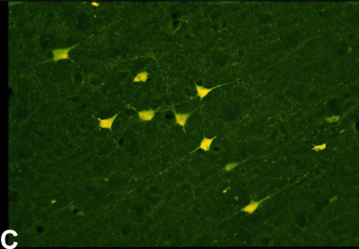
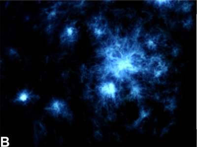
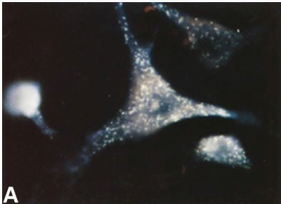
A. Retrogradely labeled neuron within the brainstem reticular formation following Fluoro-Gold injection into the thoracic ventral horn of the spinal cord.

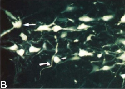
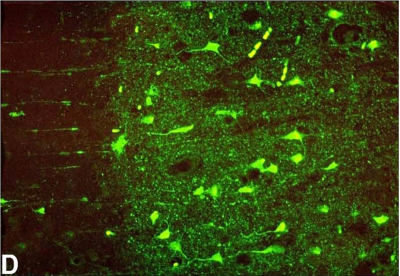
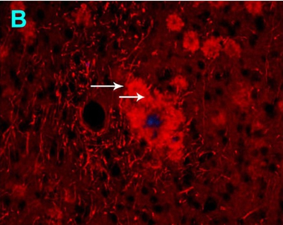
B. Neurons within the substantia nigra compacta label with Fluoro-Gold following striatal injection.

Fluoro-Ruby ®

Fluoro-Ruby

The dorsolateral striatum reveals an extensive network of axons and collaterals following the stereotaxic injection of Fluoro-Ruby into the pars reticulatta portion of the substantia nigra.

D. Dense axons and terminals are seen in the nucleus of the diagonal band following injection of Fluoro-Ruby into the dorsal medial septum.

Markers of Myelination:
Our most popular myelin stain is the gold-based brightfield stain, Black Gold II. After brief immersion of tissue sections in the Black Gold II staining solution, large bundles of myelinated fibers appear brown, while fine individual myelinated fibers appear black. In addition to revealing normal myelination, various myelin pathologies including fragmentation, edema and demyelination can be detected. Euro-Glo is a recently developed fluorescent Europium chelate that will stain myelin a pink color under UV illumination. Fluoro-Jade B and Fluoro-Jade C will also stain amyloid plaques.

Black Gold ® II

Examples of normal and pathological myelin stained with Black Gold II:

B. Black Gold II results in detailed staining of individual myelinated fibers in the dentate gyrus region of the rat hippocampus. 

E. Black Gold II staining of the rat hippocampus following exposure to kainic acid reveals both edematous and fragmented myelinated fibers.

Euro-Glo ®

Euro-Glo for the localization of myelin and amyloid plaques:

A. Euro-Glo staining of an aged AD/Tg mouse brain reveals both myelinated fibers as well as amyloid plaques in the hippocampus.

C. Euro-Glo staining of the rat parietal cortex reveals both deep radial and superficial parallel (arrows) myelinated fibers.

Localization of Neuronal Degeneration: Fluoro-Jade B and Fluoro-Jade C
Both Fluoro-Jade B and Fluoro-Jade C are used primarily to localize neurons undergoing irreversible degeneration in brain tissue sections. Both tracers produce high contrast and resolution labeling of degenerating neurons and sometimes adjacent hypertrophied astrocytes. They will also label amyloid plaques if present. Both dyes are excited by blue light and emit green light. Fluoro-Jade C is somewhat more commonly used than Fluoro-Jade B, although both tracers have their proponents.

Fluoro-Jade ® B and Fluoro-Jade ® C

Markers of neuronal degeneration:

C. Following exposure to MDMA, Fluoro-Jade B positive neurons, proximal dendrites and axonal terminals are seen in the piriform cortex.

D. Fluoro-Jade C also stains degenerating neurons, dendrites and axon terminals in the cingulate cortex following exposure to kainic acid.

Markers of AD Pathologies:
Histo-Chem offers four different tracers capable of labeling the pathologies associated with Alzheimer’s disease in tissue sections from afflicted humans or from APP/PS1 transgenic mice. These specific tracers are Amylo-Glo, Fluoro-Jade C, Euro-Glo, and HQ-O.

Amylo-Glo was developed specifically to localize amyloid plaques in AD/Tg mice and to localize both plaques and tangles in human brain tissue sections.

As previously mentioned, in addition to staining degenerating neurons, Fluoro-Jade C will also label amyloid plaques; and in addition to staining myelin, Euro-Glo will also stain amyloid plaques.

The most recently developed of the markers of AD pathology is HQ-O, which is specific for amyloid plaques and leucocytes, when present. It is thought to be bound to the zinc contained in these structures.

Amylo-Glo ®

Localization of amyloid plaques with Amylo-Glo: Cortical amyloid plaques from aged AD/Tg mouse labeled with Amylo-Glo.

B. High magnification view of amyloid plaques shows both dense core and fibular components.

C. Survey view of an entire aged AD/Tg mouse brain section is suitable for both qualitative and quantitative analysis.

Fluoro-Jade ® B and Fluoro-Jade ® C:
As previously mentioned, in addition to staining degenerating neurons, both Fluoro-Jade B and Fluoro-Jade C will also stain amyloid plaques in brain sections from AD/Tg mouse models of Alzheimer’s disease as well as from humans with the disease.

Euro-Glo ®:
Also previously mentioned was the observation that in addition to staining myelin in frozen cut tissue sections, Euro-Glo also labels amyloid plaques after several days of incubation in the staining solution.

Fig B illustrates an amyloid plaque in the hippocampus of an AD/Tg mouse model of Alzheimer’s disease. Arrows indicate red globular structures within a large central plaque with a blue central core. Four smaller plaques are at upper right, while at the left neurite-like structures are seen.

HQ-O:
Incubation of tissue sections from the brain of an AD/Tg mouse model of Alzheimer’s disease in a HQ-O solution results in the bright and specific labeling of amyloid plaques. The labeling has been attributed to zinc bound to the plaques.

Fig A is a survey view of a section of the cortex of a one year-old APP/PS1 mouse (pial surface at upper left).

Box seen at the left is shown in Fig B under higher magnification to show fibular appearance of labeling.